Achilles Tendon Rupture
Opublikowano dnia 15th Oct 2016 / Opublikowano w: Kostka

What is the Achilles Tendon?
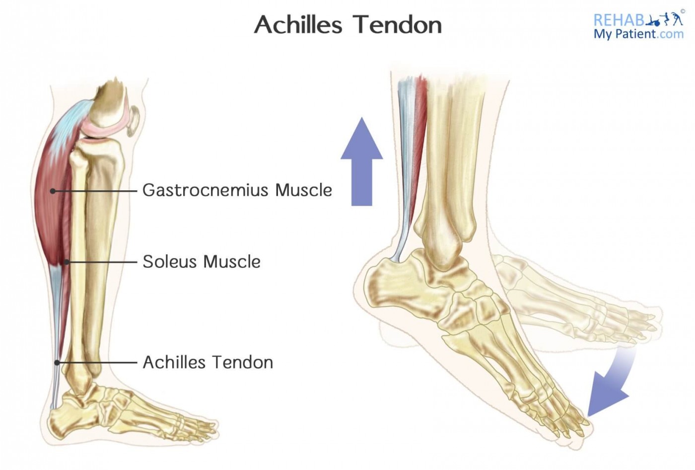
The Achilles tendon is an important structure in the back of the leg above the heel bone (calcaneus). The tendon connects the heel bone with the calf muscles (known as the gastrocnemius and soleus) and bends the foot downwards away from your shin (known as plantar flexion of the foot).

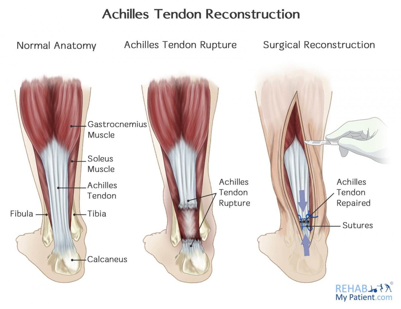
A partial or complete tear of the Achilles tendon is called an Achilles tendon rupture. The difference between partial and complete tear depends on whether the tendon is partially torn but still connected to the calf muscles (partial tear) or completely torn with no connection between the calf muscles and the heel bone (complete tear).
Mechanism of Injury
The Achilles tendon can be ruptured when high stress or force is applied on it during activities such as football, basketball, tennis or running. The rupture occurs due to the demand of a high-impact push off from the foot. Sometimes the mechanism of injury can be trivial, with patients reporting they just lunged for the ball, or turned to run to the back of the tennis court. Another less common injury is the deep cut of the tendon from direct force e.g. during a road traffic accident.
The tendon will usually rupture about 2-6cm above the heel bone. A diagnosis is often made by your doctor or therapist feeling around the site of the tendon for any obvious gaps and swelling. Also they may perform a test where you lie on your front, with your knee bend, and they squeeze your calf looking for any foot movement (if your foot moves, this is a good sign, and means your tendon is intact, but no movement is likely to indicate a full rupture). Sometimes an MRI scan is required to check the tendon in more detail.
Signs & Symptoms
· When injury occurs the patient may hear a snap or feel a very sharp pain in the back of the lower leg
· This will often drop the patient to the floor
- Inability to push off the ground
- Inability to stand on toes
- Severe limp
Surgical Treatment
Surgery is often recommended in cases of full rupture. The operation to surgically repair the tendon involves sewing of the two parts of tendon. If needed, the surgeon may use an additional tendon graft.
After surgery, a plaster cast, brace, or post-surgical boot is needed for protection and promotion of healing.
Manual therapy is also recommended to reduce inflammation, improve ankle mobility and improve strength to the foot and ankle post-surgery.
Important Information After Achilles Rupture:
- Get yourself to hospital if you suspect an Achilles rupture.
- If you are unable to weight bear through your leg and you also heard a pop or felt your Achilles “go”, get yourself to hospital.
- Surgery may or may not be performed, and it will often depend on the hospital you are seen at, the consultant who looks at your injury, or whether you have private medical insurance.
- If you are placed into a cast instead of having surgery, your ankle will be very stiff when you get out of the cast. So it is important to do plenty of ankle exercises and see a physiotherapist or manual therapy practitioner who can rehabilitate you.
- You may also be provided with a heel lift to bring the Achilles tendon closer.
- Rest, Ice and Elevate your foot when you come out of the cast to allow healing of the tendon.

Conservative Treatment
Conservative treatment involves rest in order to allow the tendon to heal naturally. A plaster is used for approximately 8 weeks aiming at protection of the tendon during the healing period. The plaster is positioned with the foot pointing downwards to avoid stress on the tendon, and to shorten the space for the tendon to heal.
Manual therapy is crucial in the rehabilitation stage immediately after plaster removal, as the foot can be very stiff, and mobility of the ankle must be improved as soon as possible. There may also be swelling around the foot and ankle. Treatment will work on reducing the inflammation and swelling, improving foot and ankle mobility, and improving muscular strength to the foot.
Zapisać się
Zarejestruj się już teraz, aby skorzystać z bezpłatnego okresu próbnego!
Zacznij korzystać z Rehab My Patient już dziś i zrewolucjonizuj proces przepisywania ćwiczeń, aby zapewnić sobie skuteczną rehabilitację.
Rozpocznij 14-dniowy bezpłatny okres próbny