Rectus Capitis Lateralis
Opublikowano dnia 28th Jul 2020 / Opublikowano w: Głowa

General information
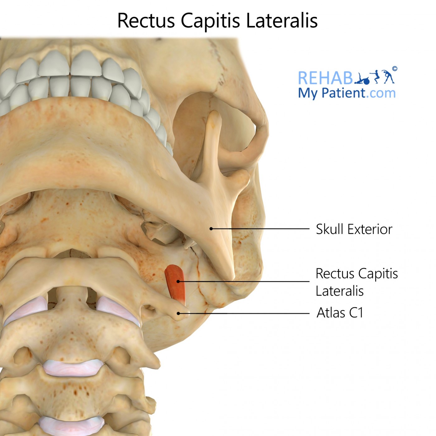
The rectus capitis lateralis is a flat, short muscle that rises from the upper surface in the transverse processes of the atlas. It is inserted into the underneath surface in the jugular process for the occipital bone.
Literal meaning
Straight muscle of the head at the side.
Interesting information
Some of the most frequent types of injuries to athletes are those of an acute strain or sprain in the musculature of the neck, in addition to any soft-tissue contusions. Strains are injuries to the muscles that occur whenever the muscle is overloaded or stretched. Sprains are when the ligaments and capsular structures that connect the vertebrae and cervical facet joints become damaged. It may be quite difficult to differentiate between the two injuries, which is why they often occur at the same time. Symptoms may be described as stiffness in the neck, muscle spasms in the affected area, tenderness and reduced range of motion of the head and neck. Treatment of an injury to this muscle include icing the affected area, isolating movement of the area, rest and massage therapy. NSAIDs, such as ibuprofen, may be taken to address pain and reduce inflammation.
Origin
Superior margin for the transverse process in the atlas.
Insertion
Jugular process for the occipital bone.
Function
Assists weakly with lateral flexion for the head.
Postural muscle that aids in monitoring the position and movement in the head.
Nerve supply
Cervical ventral rami branches C1-C2.
Blood supply
Muscle receives its blood from ascending cervical artery, which comes from a small branch in the inferior thyroid artery out of the trunk of the subclavian artery.
Vertebral artery branches.
Occipital artery passing to the lateral aspect.

Relevant research
This study aimed to collect data on the muscle stiffness of the four rectus capitis muscles, to gain a better understanding of their role in stabilising the atlanto-occipital joint. The study found that rectus capitis lateralis muscles failed at a significantly higher level of load and strain than the other three pairs of muscles. The passive stiffness of rectus capitis lateralis and anterior muscles was higher than the other muscle pairs. The authors concluded that the anatomic location and high level of passive stiffness of the rectus capitis lateralis muscles means they would normally facilitate the atlanto-occipital joint congruence. They advise that diagnostic and treatment procedures that apply force to the upper cervical spine should be adjusted accordingly with the patient’s age, gender and history of previous injuries to avoid overstretching the rectus capitis muscles.
Hallgren, R. Injury Threshold of Rectus Capitis Muscles at the Atlanto-occipital Joint. Journal of Manipulative and Physiological Therapeutics. 2017 Feb; 40 (2): 71-76.
Rectus capitis lateralis exercises
Side neck stretch
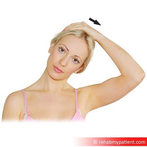
Sit up straight in a solid based and straight-backed chair. Relax the left hand into the lap while the right hand begins to reach over the top of the head. Allow the right hand, with fingers closed, to lay on the left side of the head just above the left ear. Gently press the head to the right side as if to press the right ear to the right shoulder. Hold this position for 10-15 seconds, then slowly release it back up to the starting position. Switch hands and repeat to the other side. This may be done two to three times a day.

Zapisać się
Zarejestruj się już teraz, aby skorzystać z bezpłatnego okresu próbnego!
Zacznij korzystać z Rehab My Patient już dziś i zrewolucjonizuj proces przepisywania ćwiczeń, aby zapewnić sobie skuteczną rehabilitację.
Rozpocznij 14-dniowy bezpłatny okres próbny