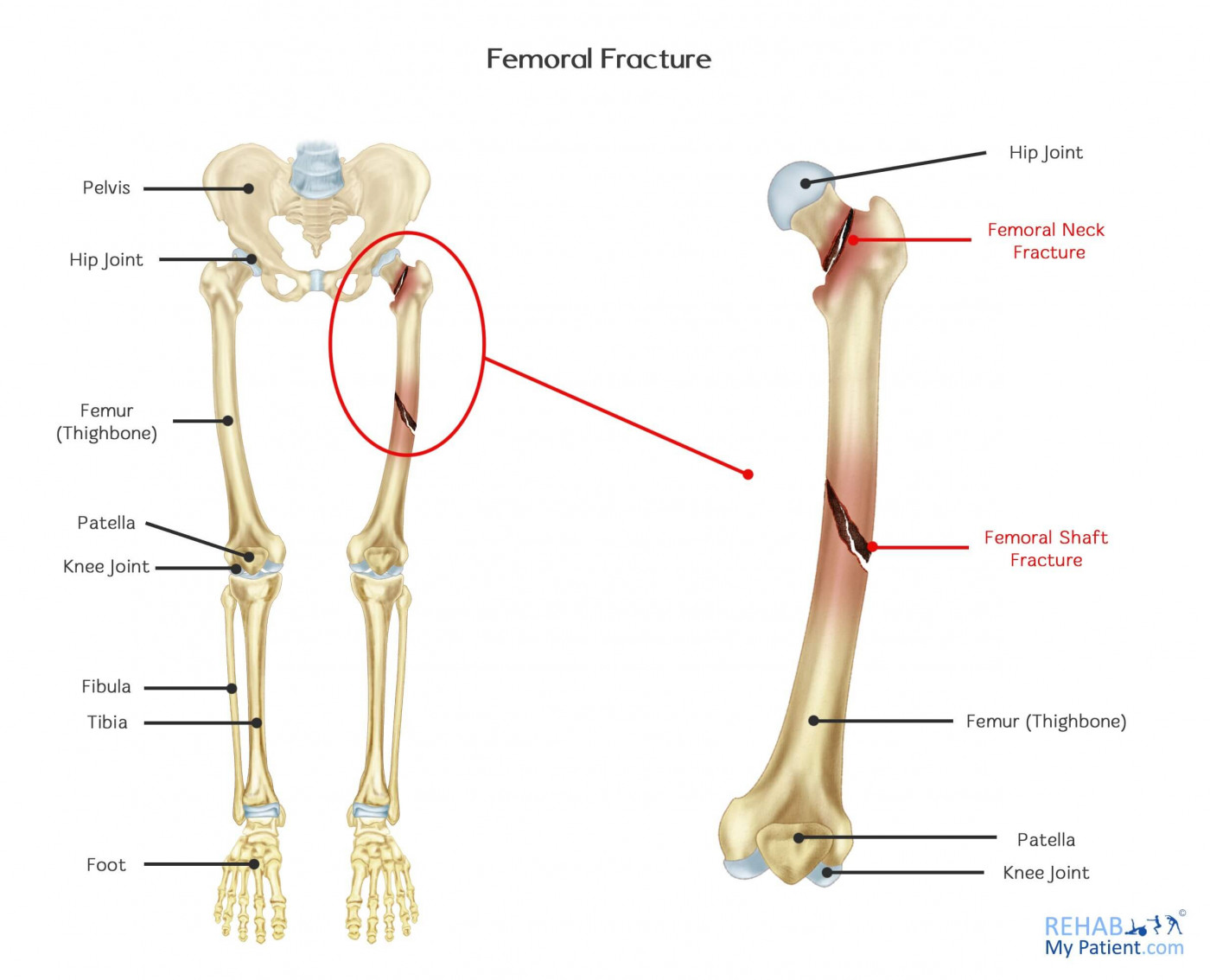
The thighbone (femur) is the longest bone found in the body. Since the femur is so strong, it will take a great deal of force to break this bone. A car crash is one of the main causes of a femoral fracture. The straight, long part of the bone is referred to as the femoral shaft. Whenever a break occurs anywhere along the length of the bone, it is referred to as a femoral shaft fracture.
A “Broken hip” could also be referred to as a femoral fracture, so it is worth discussing with your doctor where the fracture is located. Often in these cases the fracture is located at the top of the femoral shaft, where the ball connects to the neck of the femur. It is also possible to fracture the femur at the neck and this is called a femoral neck fracture.
Femoral Fracture Anatomy
The femur is the long bone located in the thigh. They have an exceptional supply of blood going to them. It is because of this and the protective surrounding muscle that the shaft doesn’t easily fracture. When fracture does occur, sometimes the force can be excessive enough to cause displacement in which case the bone dislocates from its correct position.
The leg is a lower limb of the body supporting the body when in a standing position. It provides the body with the ability to jump, walk, run and various other movements. It extends from the ankle all the way up to the hip joint, which has the largest bone within the body, the femur.

Femoral fractures are common in high trauma incidents such as car crashes or falling from a height. But the population most likely to suffer with femoral fractures are the elderly, especially elderly ladies. This is because the neck of the femur is prone to osteoporosis (bone thinning). A simple fall from tripping over a curb or pavement slab can be enough to cause a fall and fracture the femur.
How to Treat a Femoral Fracture:
- Intramedullary Nailing
The method the majority of surgeons tend to use for treating fractures is that of intramedullary nailing. During the procedure, a metal rod that is specially designed will be inserted into the narrow femur canal. The rod passes along the fracture to make sure it stays in position.
An intramedullary nail is inserted into canal at either the knee or the hip through a small incision. The nail will be screwed in at either end, which help to keep the nail and the bone where they need to be in the healing process. The nails are often made from titanium. They come in an array of different lengths and diameters to accommodate the majority of femur bones.
- Plates and Screws
During this particular operation, all of the bone fragments are repositioned back to where they are supposed to be. They are held in position with specialized screws and metal plates that affix to the outer part of the bone. Screws and plates are generally used when intramedullary nailing isn’t possible, such as that of fractures extending into the knee and hip joints.
Tips:
- Leg motion is encouraged during the early part of the recovery period. Following the instructions provided from the doctor is important to avoid any future problems with applying weight too soon.
- Depending on your particular case, you might not be able to put weight on the leg until healing has begun. For others, the patient can put more weight on the leg following the surgery. It depends on the surgery and the recommendations of the surgeon.
- When you start walking again, you will probably need to use a walker or crutches for additional support.
- Since you are probably going to lose some muscle strength at the site of the injury, you will need to begin an exercise regime during the healing process.
- Physical and manual therapy helps to restore joint motion, muscle strength and flexibility, and it should be commenced as soon as possible following surgery. Adhering to an exercise program will also be important.
Zapisać się
Zarejestruj się już teraz, aby skorzystać z bezpłatnego okresu próbnego!
Zacznij korzystać z Rehab My Patient już dziś i zrewolucjonizuj proces przepisywania ćwiczeń, aby zapewnić sobie skuteczną rehabilitację.
Rozpocznij 14-dniowy bezpłatny okres próbny