Mylohyoid
Opublikowano dnia 27th Jul 2020 / Opublikowano w: Szyja

General information
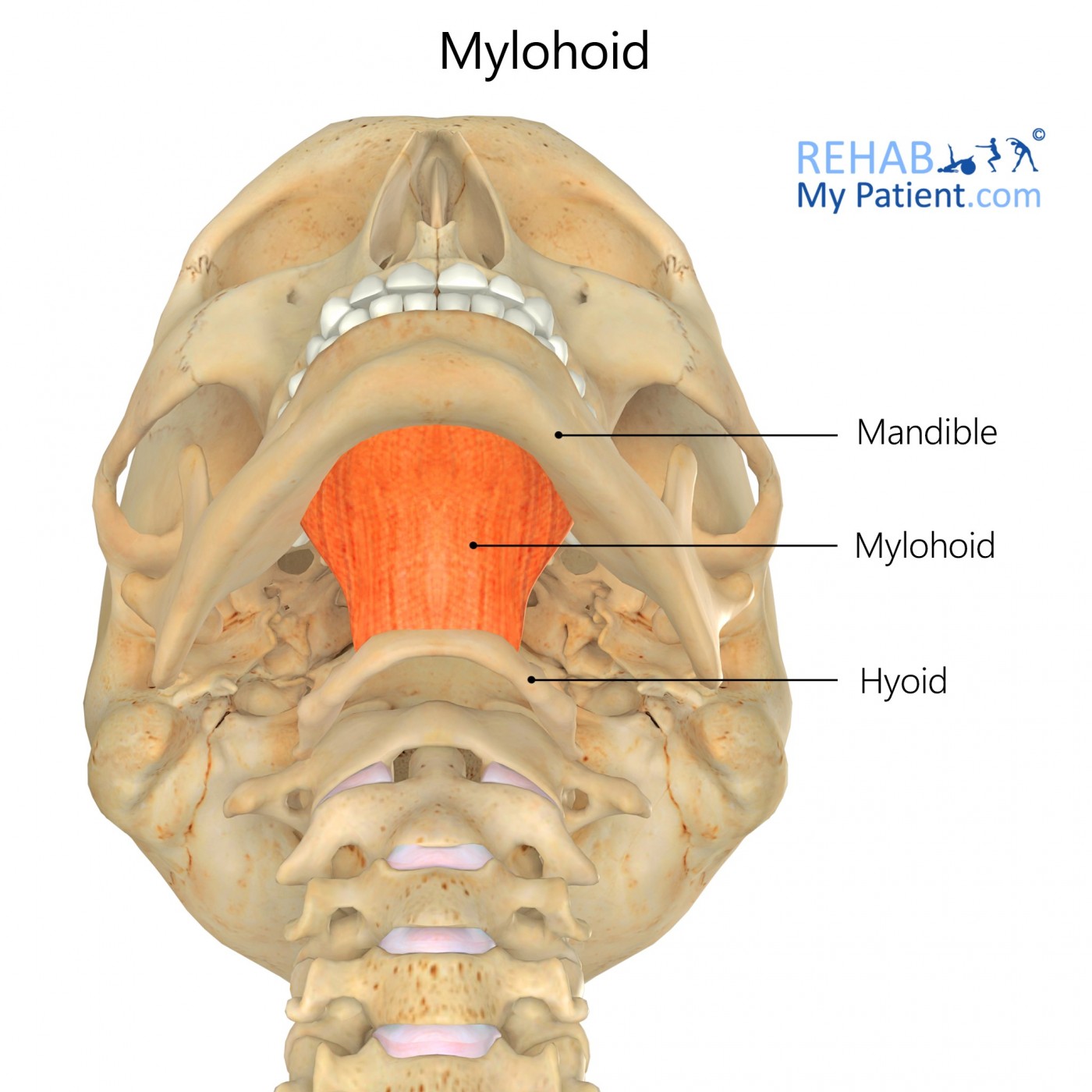
Mylohyoid is the muscle that begins at the jaw and extends toward the hyoid bone.
Literal meaning
The U-shaped grinder.
Interesting information
Mylohyoid is a triangular-shaped skeletal muscle located between the jawbone and the hyoid bone that aids in elevation of the oral cavity floor during swallowing.
The mylohyoid is part of a group of muscles called the suprahyoids. Lymphangiomas are soft masses that may form on either side of the neck in either the suprahyoid or the infrahyoid region. These malformations of the lymphatic system normally present themselves before the age of two, but may occur at any time due to trauma, obstruction of lymph through the lymph nodes, or inflammation. Other causes are alcohol use during pregnancy, viral infection during pregnancy, or genetic disorders, like Down or Turner Syndrome. Treatment includes surgical removal, lasers, and sclerotherapy but, if not causing any symptoms, the masses may be left alone since they are benign. In rare cases, complications with swallowing, breathing, leaking of lymph fluid, slight bleeding, or infections of the neck may arise. If a parent has a child with lymphangioma, it is possible the cause is genetic. In this case, the parents should consider seeking genetic counselling before having more children.
Origin
Mylohyoid line (mandible).
Insertion
Body of hyoid bone and median raphe.
Function
Elevation of oral cavity floor.
Elevation of the hyoid.
Elevation of the tongue.
Depression of the mandible.
Nerve supply
Mylohyoid nerve.
Blood supply
Mylohyoid branch of inferior alveolar artery.

Relevant research
During cadaver dissections, it is often common to find variations of nerves, arteries, or veins throughout the body. A routine dissection led to the finding of a new anomalous pattern of the mylohyoid nerve and its branches, originating from the mandibular trunk. Awareness of this anomaly is important for surgeons to know about when approaching the inferior alveolar nerve.
Kumar, S, Kumar C, Bhat, S, Kumar A. (2011). “Anatomical study of the unusual origin of a nerve to the mylohyoid muscle and its clinical relevance”. British Journal of Oral and Maxillofacial Surgery. 49:5, e14-e15.
Case studies show that intramuscular hemangioma of the neck and head is uncommon. These types of hemangiomas are most frequently located in the masseter or the trapezius muscle, but in very rare cases they are located in the mylohyoid. The tumours are located deep within the muscle and may either be soft and diffuse or firm and localised. Treatments may include surgical excision, steroids, radiation, or cryotherapy (freezing).
Lee, J, Lim, S. (2005). “Intramuscular hemangiomas of the mylohyoid and sternocleidomastoid muscle”. Auris Nasus Larynx. 32:3, 323-327.
Mylohyoid exercises

Kiss the Sky
To begin, tilt the head and look toward the ceiling. Pucker lips to mimic a kissing motion and extend lips as far as possible. Hold the position for ten seconds, release, and lower the head. Repeat the exercise five times a day.
Tongue touches
Look upward toward the ceiling and open the mouth as wide as possible. Try to touch the chin with the tongue. Hold the position for five seconds and return to the starting position. Perform the exercise three times a day.
Zapisać się
Zarejestruj się już teraz, aby skorzystać z bezpłatnego okresu próbnego!
Zacznij korzystać z Rehab My Patient już dziś i zrewolucjonizuj proces przepisywania ćwiczeń, aby zapewnić sobie skuteczną rehabilitację.
Rozpocznij 14-dniowy bezpłatny okres próbny