Omohyoid (neck)
Opublikowano dnia 29th Jul 2020 / Opublikowano w: Szyja

General information
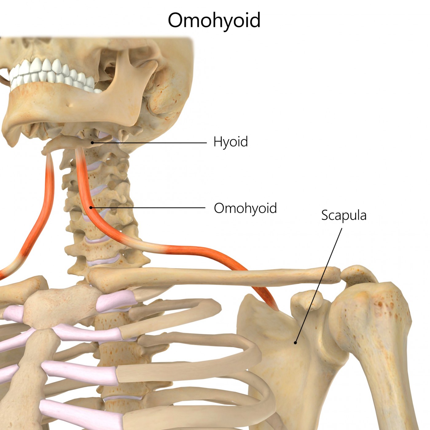
Omohyoid is a muscle of the cervical wall extending from the hyoid bone to the scapula.
Literal meaning
The muscle of the shoulder and hyoid bone.
Interesting information
The omohyoid muscle is situated in the neck and is comprised of two distinct sections – the inferior belly and the superior belly. The two sections are separated by an intermediate tendon.
The inferior belly of omohyoid has its origin at the superior edge of the scapula and crosses through the scapular notch. There is considerable anatomical variation in the width of omohyoid attachment to the scapula; it can range from just a few millimetres to nearly three centimetres.
The superior belly of this muscle follows a nearly vertical trajectory along the sternohyoid. It eventually inserts at the inferior surface of the hyoid body.
The intermediate tendon which separates the inferior belly from the superior belly varies in length and is sheathed in cervical fascia which attaches at the first rib and clavicle. This attachment is what allows the muscle to maintain its obtuse angular shape.
The primary function of omohyoid is to depress the hyoid bone which aids in the actions of chewing and swallowing. Injury to the omohyoid muscle is particularly common among bodybuilders and those involved in weight bearing exercise. Pain is often referred to the omohyoid when there is damage to the surrounding structures such as neck whiplash injury following an automotive accident.
Origin
Inferior belly: superior edge of the scapula (scapular notch) and superior transverse scapular ligament.
Insertion
Superior belly: inferior surface of the hyoid body.
Function
Depresses the hyoid bone and aids in raising cervical fascia during inhalation.
Nerve supply
Inferior belly: ventral ramus (C2, C3).
Superior belly: ventral ramus (C1).
Blood supply
Infrahyoid artery.

Relevant research
Venous entrapment is a very serious and potentially life-threatening condition. Entrapment of the internal jugular vein has been shown to sometimes be caused by the omohyoid muscle. However, additional research on patho-physiological aspects of omohyoid venous compression needs to be completed.
Gianesini S, Menegatti E, Mascoli F, Salvi F, Bastianello S, Zamboni P. “The omohyoid muscle entrapment of the internal jugular vein. A still unclear pathogenetic mechanism”. Phlebology. 2013 May 16.
Serial photography, real-time ultrasonography, computed tomography, and videography have been used to observe the transient tenting of sternomastoid in patients with omohyoid sling syndrome. Analysis of these recordings have pointed to specific anomalies in omohyoid which bring about this pathology.
Wong DS, Li JH. (2000). “The omohyoid sling syndrome”. Am J Otolaryngol. 21(5):318-22.
Omohyoid exercises


Neck extension and flexion
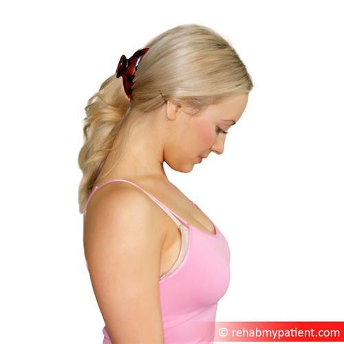
General neck toning exercises can strengthen omohyoid and the surrounding muscular structures. Begin by standing comfortably with your feet shoulder width apart. Now slowly arch your neck forward. Don’t simply drop your chin to your chest but rather imagine that you are curling your neck over a small object near your throat. Then slowly arch your neck backwards using the same technique. It is imperative that your movements are slow and fluid to prevent injury to your neck. Your shoulders and entire upper body should be relaxed and remember to breathe deeply throughout the exercise. Repeat the entire motion a total of ten times.
Zapisać się
Zarejestruj się już teraz, aby skorzystać z bezpłatnego okresu próbnego!
Zacznij korzystać z Rehab My Patient już dziś i zrewolucjonizuj proces przepisywania ćwiczeń, aby zapewnić sobie skuteczną rehabilitację.
Rozpocznij 14-dniowy bezpłatny okres próbny