Semispinalis Capitis
Opublikowano dnia 29th Jul 2020 / Opublikowano w: Szyja

General information
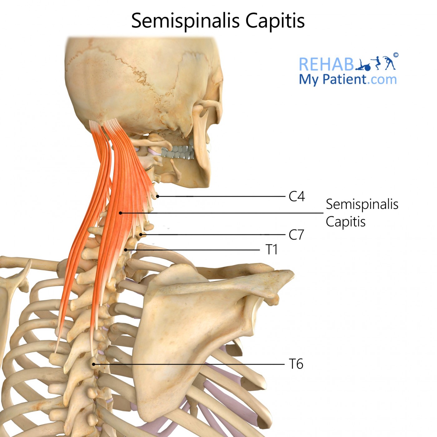
The Semispinalis capitis is found at the upper back portion of the neck, medial to the longissimus, capitis and cervicis and deep to the splenius. This muscle is part of the transversospinales muscle group. The muscle arises from a series of tendons along the tips of the transverse processes for the upper six to seven of the cervical and thoracic vertebrae, as well as from the articular processes for the three cervical vertebrae above C4-C6.
Tendons unite to form a broad muscle, which passes up and is inserted between inferior and superior nuchal lines for the occipital bone. Nestled deep within the trapezius muscle, it can be palpated as a firm, round muscle mass that is lateral to the cervical spinous processes. These muscles are innervated from the dorsal rami in the cervical spinal nerves.
Literal meaning
Half thorn head.
Interesting information
Most of the time, the Semispinalis capitis muscle is injured from a blow to the head or whiplash from being in a car accident. The most often felt symptom of an injury to this area is the feeling of tenderness in the back of the head and neck. Other common symptoms of injury are numbness in the scalp, pain in the temple shooting to the eye, headaches, and pain in the upper part of the neck and into the head. Treatments often include rest, isolation of the head and neck in a cushioned apparatus, taking anti-inflammatory medications to reduce pain and swelling, as well as physical therapy exercises during rehabilitation.
Origin
Transverse processes tip of C7 to T6.
Articular tubercle of C4-C6.
Insertion
Medial part of the space between inferior and superior nuchal lines for the occipital bone.
Function
Extension of the head.
Weak rotation for the head to the opposite side.
Nerve supply
Dorsal rami for the thoracic and cervical spinal nerves C2-T6.
Blood supply
Lateral cervical portion for the muscle is supplied from the deep cervical artery arising from the costocervical trunk related to the subclavian artery.
Occipital artery descending branch forming an anastomosis within the deep cervical artery.

Relevant research
Studies indicate that injury produces changes in the plasticity for various neuronal structures responsible for amplification of exaggerated and nociception pain responses. Consistent evidence exists of hypersensitivity in the central nervous system relating to sensory stimulation in chronic pain following a whiplash injury. Tissue damage tends to be the leading cause of central hypersensitivity. Different mechanisms co-exist and underlie when it comes to chronic whiplash conditions. Predominantly neuropathic pain components are related to higher disability and pain levels.
Davis CG. Mechanisms of chronic pain from whiplash injury. J Forensic Leg Med. 2013;20(2):74?85. doi:10.1016/j.jflm.2012.05.004
Semispinalis capitis exercises
Neck retraction exercises
Sit on a soft surface or chair and engage the core to help stabilise the spine. Brace the posterior muscles in your neck to bring the head backward into a stable position while having the chin slightly downward. Hold the position briefly. Relax and repeat. Start with one set of five repetitions and work your way up to ten repetitions in total.
Zapisać się
Zarejestruj się już teraz, aby skorzystać z bezpłatnego okresu próbnego!
Zacznij korzystać z Rehab My Patient już dziś i zrewolucjonizuj proces przepisywania ćwiczeń, aby zapewnić sobie skuteczną rehabilitację.
Rozpocznij 14-dniowy bezpłatny okres próbny